
Magnetic Resonance Laboratory
Transport phenomena in complex materials
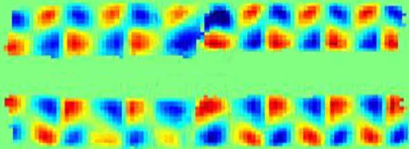
Magnetic Resonance Imaging (MRI) is a noninvasive experimental technique which has found broad application in clinical medicine and is a maturing method for studies in engineering and physics. Our group uses instruments that allow MRI to be applied with a resolution of 10 µm over 10 mm diameter samples. MRI applied on this scale requires more sample specific tailoring of the pulse sequences and is more often termed Magnetic Resonance Microscopy (MRM) to distinguish it from medical scale imaging.
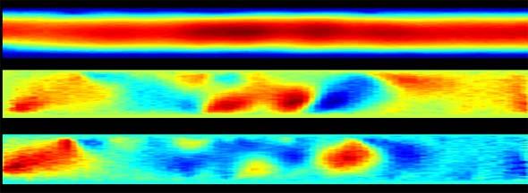
Our work is concerned with furthering application of MRM methods in the study of transport phenomena and material characterization. We use pulsed gradient spin echo (PGSE) techniques to measure velocity and effective diffusion, e.g. dispersion. The ability to spatially resolve velocity and diffusion fields allows for transport visualization. Application of PGSE methods without spatial resolution provide the scale dependent statistics of motion over the entire sample, rendering it a powerful technique for studying anomalous transport phenomena. The research program seeks to both elucidate new transport phenomena in complex systems and develop MRM methods for new applications.
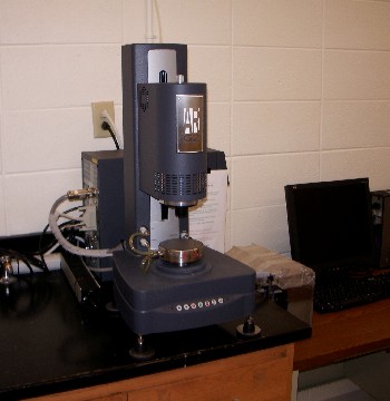
Rheology: We have a top of the range TA instrument rheometer that can measure both shear stress under controlled shear rate and shear rate under controlled shear stress conditions as well as a range of oscillatory experiments. We also use a RheoNMR insert that allows rheology measurements to be made using the NMR spectrometer
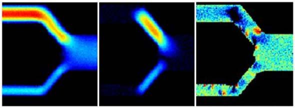
Simulations: We use Comsol to simulate simple fluids (Newtonian, power-law) in simple 2D structures
(capillary networks, Couette cells, cone and plate cells) and compare to our experimental
results from more complicated fluids (colloidal suspensions, blood, viscoelastic).
We also collaborate with an expert in the Lattice-Boltzmann simulation technique.
This method is more challenging to use but yields simulations of richer and more complex
2D porous media structures.
Bruker AVANCE250
Micro5 Probe
Maximum Gradient: 3 T/m 60A
Max Sample Dimension: 1 cm
Micro2.5 Probe
Maximum Gradient: 1.5 T/m 60A
Max Sample Dimension: 3 cm
Diff30 Probe
Maximum Gradient: 17 T/m 60A
Max Sample Dimension: 1 cm
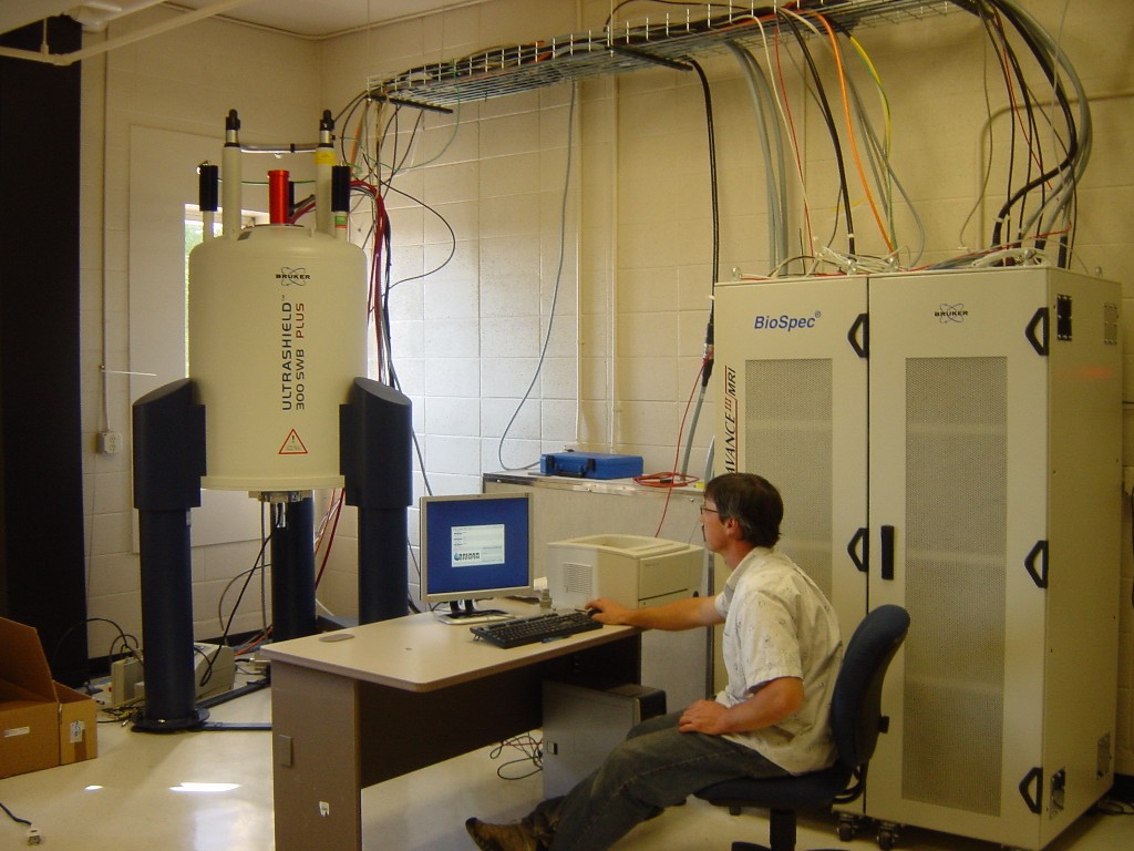
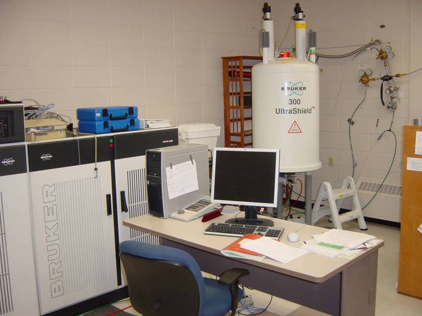
Bruker AVANCE300
Micro2.5 Probe
Maximum Gradient: 1.5 T/m 60A
Max Sample Dimension: 3cm
SuperWideBore Probe
Maximum Gradient: 0.37 T/m 100A
Max Sample Dimension: 6cm
We have two state-of the art NMR/MRI instruments. These instruments are invaluable in the medical field for diagnosis of soft tissue damage and used routinely by biochemists to probe molecular structure. We use these instruments to obtain images as well as velocity and diffusion measurements in all the important systems described above. Magnetic Resonance techniques have the unique capability of allowing transport studies in opaque systems and can make non-invasive measurements that do not disturb the very flow in which we are interested.
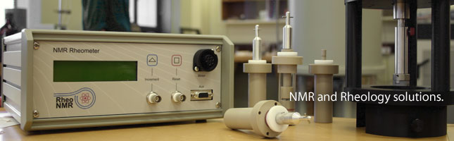
RheoNMR
Rehometer that operates inside the NMR spectrometer. Couette and cone and plate cells availabel. Max rotation rate: 14 Hz
Rheometer
TA Instruments
We have a top of the range TA instrument rheometer that can measure both shear stress
under controlled shear rate and shear rate under controlled shear stress conditions
as well as a range of oscillatory experiments.
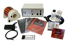
Earths Field MRI
Portable MRI Instrument that uses the earth's magnetic field. Used for training and undergraduate research projects.
The Magnetic Resonance Lab is situated in the College of Engineering at Montana State University[BROKEN LINK] in the spectacular location of Bozeman in the heart of the Rocky Mountains of the USA.
Transport + Rheology + Simulations + MRI + NMR
Ceramics and Foams
These modern materials are invaluable to the alternative energy industry, food industry and medical industry. Our ability to increase the range of application of fuel-cells, or design next generation filtration systems for industry and medicine, depends on our ability to improve our understanding of transport in the complex structures of designed porous media such as new ceramics and foams.
Colloidal Suspensions
A colloidal suspension consists of solid particles (typically less than 10 µm) that are suspended by thermal Brownian motion and do not sediment in a suspending liquid. Fluids as diverse as paints and blood are classified as colloidal suspensions. They demonstrate fascinating shear-thinning, shear-thickening, particle migration and deposition effects – many of which are still not fully understood. Transport of colloidal suspensions under shear forces plays an elemental role in industry and biology. In natural systems, the dynamics of cellular bacteria in microbiology, red blood cells in physiology, and colloidal contaminants in earth formations impact the system function and transport. Understanding colloidal dynamics is also required for design of products such as drug delivery agents and microfluidic devices. Our lab can manufacture model colloidal suspensions of solid oil filled core shell particles suspended in water and stabilizing chemicals. The model particles allow magnetic resonance techniques to access transport features of the suspending liquid separate from the suspended particles.
Biofilms
Biofilms are microbial colonies that grow on surfaces. Their significance has encouraged a flood of recent research in areas as diverse as environmental bioremediation and biomedical applications. Biofilms are responsible for oral plaque and the persistent infections in catheters, medical implants and lungs. There is still not much known about the mechanics of and diffusion within the viscoelastic biopolymeric gel surrounding the bacteria and hence the effectiveness of agents to penetrate from the bulk fluid, but it is known that biofilms play a significant role in the resistance of bacteria to antibiotic treatment. The shear stresses experienced during growth may play a role in explaining antibiotic resistance and structural survival even during high and turbulent flow. While much is known about the growth state of free bacteria, much less is known about the structure-function relationships when cells are in the biofilm state. We have a strong collaboration with the Center for Biofilm Engineering and access to their expertise and equipment. We have used a wide range of magnetic resonance techniques to study the macroscale impacts of biofouling of fluid transport in medical, industrial and environmental materials and we study the fundamental rheological properties of the biofilm slime itself.
Porous Media
The underlying physics of transport in porous media is relevant to the aforementioned ceramics and foams as well as a plethora of other applications like industrial packed bed chemical reactors, transport in gels and tissues for drug delivery, storage of supercritical CO2 in natural formations and in sub-surface transport of environmental contaminants in the earth’s subsurface. Many porous media of interest have structures that generate complex dynamics which can be modeled by fractal and percolation theory concepts. MR methods provide unique data for scale dependent transport in porous media since most are opaque and not amenable to analysis by other methods. Modeling the impact on transport dynamics of biological and chemical reactions in porous media presents a significant challenge for design of environmental remediation strategies and new materials.
Gels
Polymeric gels are of major importance in technology and nature. The transport of particles and solvents in gels is a major feature of drug delivery strategies using nanoparticles, where gels are a model “tissue” and for drug delivery via gel swelling. Understanding the interplay between the underlying gel structure and transport processes will enhance these applications. Gels are also found in natural systems, such as the biofilms mentioned above, and their role in biological function an outstanding area of research interest.
Learn about the techniques we use to study these systems
Click on an image to view as a slideshow.


Click on an image to view as a slideshow.
Click Here for Post-Doc and Student Research Opportunites
You can read more details about us,our research projects, or see a list of our publications and presentations on this website